

Understanding the Risks: Why You Shouldn't Ignore an Impacted Tooth's Untreated Consequences
An impacted tooth is a tooth that fails to erupt fully through the gum line because it is blocked by bone, soft tissue, or adjacent teeth, and leaving one untreated can trigger a cascade of local and systemic problems. This article explains what an impacted tooth is, how impaction develops, and why early evaluation matters for preventing infection, cyst formation, root damage, and changes to bite alignment. You will learn to recognize common symptoms, understand the mechanisms behind complications like pericoronitis and root resorption, and see when extraction or surgical exposure is the recommended path. The guide also reviews recovery expectations, practical aftercare steps to reduce risks such as dry socket or spreading infection, and referral points for oral surgery consultation. Throughout, clinical terms are defined, radiographic indicators (X-ray/CBCT) are described, and action-oriented guidance is provided so you can make informed decisions about care, including local treatment options available in Lutz, FL.
What Is an Impacted Tooth and How Does It Affect Oral Health?
An impacted tooth is a tooth whose eruption is arrested by tissue or bone, creating a trapped crown or root beneath the gum; this entrapment alters normal oral anatomy and increases risk for local pathology. The mechanism begins when insufficient arch space, abnormal angulation, or delayed eruption causes a tooth—most commonly a third molar—to press against adjacent structures, creating pockets that trap bacteria and food debris. The specific benefit of early identification is that clinicians can prevent downstream complications such as infection, cystic change, and damage to neighboring teeth through targeted monitoring or timely intervention. Current research shows that radiographic assessment with dental X-rays or CBCT imaging provides the definitive anatomical detail clinicians use to plan treatment. Understanding the anatomical relationship between crown, root, gum tissue, and jawbone clarifies why an impacted tooth can affect periodontal health, occlusion, and even nerve proximity.
Which Teeth Are Commonly Impacted?
Third molars, commonly called wisdom teeth, are the most frequently impacted teeth due to evolutionarily reduced jaw size and their late eruption timing, often in late teens to early twenties. Canines—especially upper canines—are the second most common site of impaction; when a canine fails to erupt it can cause orthodontic complications, prolonged treatment time, and aesthetic concerns. Less commonly, premolars or supernumerary teeth can be impacted and disrupt occlusion or root alignment. Age and developmental stage influence prevalence: younger patients evaluated during late adolescence may benefit most from monitoring to decide between orthodontic exposure, surgical extraction, or conservative management. This distribution of hyponyms (impacted wisdom tooth, impacted canine tooth, impacted molar) highlights the range of impaction patterns clinicians encounter and informs follow-up strategies.
Further emphasizing the prevalence and anatomical factors contributing to this issue, research highlights specific reasons why third molars are so frequently impacted.
Causes of Third Molar Impaction & Extraction Frequency
Extraction of the third molar is the most frequently performed procedure in oral and maxillofacial surgery1. Owing to insufficient space between the anterior border of the mandibular ramus and the distal surface of the second molar as well as anatomical factors such as palatal impaction, the mandibular third molar has a high impaction rate2. In relation to maxillary third molars, factors such as proximity to the maxillary sinus and the presence of dense cortical bone can contribute to impaction of the third molar3.
Comparison of safety of general anesthesia and intravenous sedation during third-molar extraction surgery, MH Karm, 2024
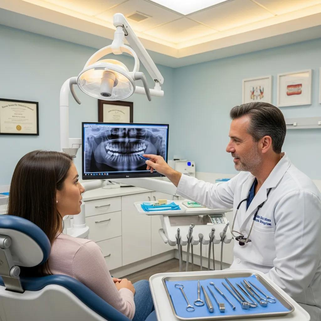
What Are the Symptoms and Signs of an Impacted Tooth?
An impacted tooth can produce a mix of local and systemic signs that range from subtle to acute, and early recognition supports timely referral for imaging and treatment. Patients often report localized pain or tenderness near the gum where eruption is obstructed, and dentists may observe swelling, erythema, or a raised operculum over a partially erupted tooth. Radiographic signs—crowding, abnormal angulation, or radiolucent areas suggestive of cysts—are frequently the first objective indicators on X-ray or CBCT. Practitioners evaluate symptom clusters alongside imaging to distinguish transient discomfort from infection or progressive complications. Recognizing these signs allows clinicians to recommend watchful waiting, prophylactic removal, or surgical exposure depending on the clinical trajectory.
The most common patient-reported and clinical indicators include:
- Localized gum swelling and tenderness: Inflamed soft tissue over the erupting crown signals bacterial trapping and irritation.
- Recurrent localized infection or bad taste/odor: Pericoronal pockets can produce chronic malodor and intermittent abscess drainage.
- Difficulty opening the jaw (trismus) or referred jaw pain: Muscle guarding and limited mouth opening often accompany acute infection.
- Visible partial eruption with a soft tissue flap: An operculum that traps food is a classic physical finding.
- Radiographic irregularities on dental X-ray/CBCT: Angulation, retained follicle, or radiolucency around the crown suggests pathology.
- Systemic signs in severe cases: Fever and swollen lymph nodes indicate spreading infection and require urgent care.
These symptoms form a practical checklist for patients and clinicians; when multiple items are present, expedited evaluation is advisable to reduce the chance of progression to more serious complications.
How Can You Identify Impacted Wisdom Tooth Infection Symptoms?
Pericoronitis—soft tissue inflammation around a partially erupted wisdom tooth—typically begins with localized throbbing pain that worsens with chewing and may be accompanied by swelling and tenderness of the gum flap. As bacteria colonize the pericoronal pocket, patients may notice a persistent bad taste or drainage, and the area can fluctuate between painful flare-ups and quiescent periods, making the condition appear intermittent. Red flags include rising pain intensity, spreading facial swelling, trismus that limits oral intake, fever, or systemic malaise; these signs suggest an abscess or deeper soft tissue infection that can require urgent drainage or surgical removal. If any systemic symptoms occur, prompt clinical evaluation is necessary to prevent extension into deeper fascial spaces or systemic spread.
What Are the Risks and Complications of Leaving an Impacted Tooth Untreated?
An untreated impacted tooth can lead to several predictable complications that escalate from local inflammation to structural damage and, rarely, systemic involvement; understanding these risks helps patients weigh watchful waiting against proactive treatment. Mechanistically, a trapped tooth creates pockets that retain bacteria and food particles, leading to pericoronitis and abscess formation; chronic inflammation around the follicle can stimulate cyst development such as odontogenic cysts, which may enlarge and cause focal bone loss. Adjacent teeth face increased risk of root resorption and caries due to close contact and impaired hygiene access, while malocclusion and crowding can progress over time. In advanced or neglected cases, persistent infection can threaten the integrity of the jawbone, complicate future restorative options, and, rarely, seed systemic infection in medically vulnerable individuals.
How these complications progress depends on the lesion type and patient factors, which is why early radiographic monitoring and assessment of symptoms are critical. Clinicians often choose intervention when objective evidence of structural damage, recurrent infection, or radiographic cystic change appears, and when adjacent anatomy is at risk. After considering these risks, some patients opt for extraction or surgical management to prevent further damage; for local treatment examples, Bespoke Dental in Lutz, FL offers oral surgery services, including extractions and IV sedation as clinical options for indicated cases.
How Does Infection Develop from an Untreated Impacted Tooth?
Infection typically begins with colonization of the pericoronal pocket by oral bacteria that exploit trapped food and anaerobic conditions; this environment encourages formation of a mixed aerobic-anaerobic biofilm that provokes tissue inflammation. The inflammatory response causes localized swelling and decreased local immune clearance, allowing pathogens to progress deeper into soft tissue or the follicular space; unchecked, this can form an abscess that applies pressure to bone and adjacent tooth roots. Timelines vary: acute pericoronitis can develop over days, while cystic transformations or chronic abscesses evolve over months to years. Recognizing early signs—persistent tenderness, pus, or systemic symptoms—permits drainage, antimicrobial therapy, and definitive management that reduce risk of spread.
Lasting infection risk also connects to systemic health: patients with compromised immunity or uncontrolled diabetes face higher risk for rapid progression, which reinforces the need for prompt evaluation and individualized treatment planning.
What Damage Can Impacted Teeth Cause to Adjacent Teeth and Jaw?
Impacted teeth exert pressure on neighboring crowns and roots, a mechanical relationship that can lead to root resorption of adjacent teeth, localized bone remodeling, and increased decay risk where oral hygiene is impeded. Root resorption often appears on radiographs as irregularity of the adjacent root surface and may be asymptomatic until significant structure is lost; if resorption compromises tooth vitality, endodontic treatment or extraction may be required. Cystic lesions originating from the impacted follicle can expand within the jawbone, causing cortical thinning and displacement of teeth; in severe cases, this necessitates surgical enucleation and bone grafting. Additionally, impacted teeth can influence occlusion by creating crowding that propagates misalignment and may require orthodontic intervention if left unaddressed.
Because these structural effects are often radiographically apparent before symptoms worsen, clinicians rely on imaging to decide whether conservational monitoring or proactive removal best preserves neighboring tooth prognosis.
When Is Impacted Tooth Removal Necessary?
Removal of an impacted tooth is indicated when the risk of complication or the presence of symptoms outweighs the risks of extraction, and clinical criteria guide this decision rather than an automatic approach. Clinicians assess symptomatic indicators such as recurrent infections, intractable pain, and progressive swelling alongside radiographic markers like cystic change, root resorption of adjacent teeth, unstable angulation that threatens neighboring structures, or a follicular space suggestive of pathology. Age and overall health influence timing—younger bone heals more predictably—whereas complex anatomic relationships or nerve proximity may necessitate referral to an an oral surgeon for advanced imaging and surgical planning. The decision balances watchful waiting when asymptomatic and low-risk against early removal to prevent future morbidity.
For cases that meet referral criteria, Bespoke Dental’s oral surgery expertise and availability of IV sedation provide a practical pathway for patients seeking definitive treatment; to discuss whether removal is appropriate, call Bespoke Dental at (813) 807-4902 to schedule an evaluation with their team.
What Are the Criteria for Oral Surgery Consultation?
Patients should seek an oral surgery consultation when clinical and radiographic findings suggest a complicated extraction or increased risk of complications, including unfavorable angulation, suspected cyst or tumor, close proximity to the inferior alveolar nerve, or prior failed attempts at conservative care. Expect the consult to include a thorough medical history review, focused oral exam, and updated imaging—often a panoramic X-ray or CBCT—to map tooth position relative to teeth, roots, and neurovascular bundles. Bring any prior imaging and a list of medications and allergies to streamline planning; the surgeon will discuss anesthesia options, potential risks, and the anticipated recovery timeline. Early consultation ensures that complex anatomy and sedation needs are recognized and managed in a controlled surgical setting.
A timely oral surgery referral minimizes unforeseen intraoperative complications and enables a coordinated plan for pain control and wound management that speeds recovery and preserves adjacent structures.
How Does IV Sedation Improve Comfort During Impacted Tooth Extraction?
IV sedation offers a controlled level of conscious sedation that reduces anxiety, suppresses pain perception, and facilitates complex surgical procedures by producing deep relaxation while maintaining patient safety under continuous monitoring. For anxious patients or cases requiring extended surgical time—such as removal of multiple impacted teeth or difficult angulations—IV sedation provides comfort, amnesia for the procedure, and better operative conditions for the surgeon. Safety protocols include pre-operative assessment, intraoperative monitoring of vital signs and oxygenation, and post-anesthesia recovery oversight; these measures minimize sedation-related risks and support predictable recovery. Bespoke Dental lists IV sedation among its available approaches, which can be discussed during the oral surgery consultation to match sedation level to procedural complexity and patient needs.
Using IV sedation when indicated can improve patient cooperation and surgical efficiency, shortening the procedure and often reducing immediate postoperative discomfort.
What Is the Recovery Process After Impacted Tooth Extraction?
Recovery after an impacted tooth extraction follows predictable phases that clinicians use to counsel patients and manage expectations; following recommended aftercare reduces complications and speeds tissue healing. Immediately after surgery, clot formation within the socket initiates the first phase of healing while inflammation peaks and analgesia is most needed; over the next week soft tissue closure progresses and swelling typically subsides. By two weeks most discomfort and limited function improve substantially, and bone remodeling continues over months beneath the mucosa. Knowing the typical timeline and key aftercare steps allows patients to recognize normal recovery versus warnings that require re-evaluation.
The recovery timeline below summarizes common phases and practical aftercare actions clinicians advise to prevent complications such as dry socket or infection.
PhaseTypical TimelineKey Aftercare StepsImmediate post-op0–72 hoursRest, cold packs, prescribed analgesics, avoid spitting/using strawEarly healing3–7 daysGentle rinsing with saline after 24 hours, soft diet, maintain oral hygiene around siteIntermediate recovery1–2 weeksResume most normal activities, monitor for signs of infection, follow-up visit as scheduledBone remodeling1–3 monthsGradual bone fill of socket, resume full oral hygiene and restorative planning if needed
This phased approach helps patients prepare for typical milestones and clarifies when further care is necessary.
What Are the Recommended Aftercare Steps to Prevent Complications?
Appropriate aftercare focuses on protecting the initial clot, controlling inflammation, and preventing infection; patients should follow a structured plan to minimize risks like dry socket and delayed healing. For the first 24–72 hours apply cold packs intermittently to reduce swelling, take prescribed or recommended analgesics as directed, and avoid vigorous rinsing, spitting, or using straws that can dislodge the clot. After 24 hours gentle saline rinses several times daily help remove surface debris while preserving the clot; maintain a soft diet and avoid smoking, which dramatically increases dry socket risk. Contact your surgical team promptly if you notice worsening pain after day 3, persistent bleeding, fever, or numbness—these are red flags that require prompt reassessment.
The following concise list summarizes essential aftercare precautions:
- Protect the clot: Avoid spitting, suction, and straws for at least 72 hours.
- Control swelling: Use intermittent cold packs for the first 48–72 hours.
- Medication adherence: Take analgesics and any prescribed antibiotics exactly as directed.
- Oral hygiene: Resume gentle brushing around the site after 24 hours and rinse with saline.
These steps reduce the likelihood of complications and support timely healing; if concerns arise, contacting the treating practice ensures early management and improved outcomes.
Why Choose Bespoke Dental in Lutz, FL for Impacted Tooth Treatment?
Bespoke Dental provides focused oral surgery services in Lutz, combining specialist expertise with technology and patient-centered protocols suited to impacted tooth management. The practice lists oral surgery including wisdom teeth extraction and other extractions, IV sedation availability, and advanced dental imaging as core treatment capabilities; these services support safe planning and execution for complex extractions. Board-certified oral surgeons, including Dr. Tarik Elmohd, bring surgical credentialing and experience that translate to meticulous pre-operative planning, risk mitigation for nerve proximity, and coordinated post-op care. For patients seeking local solutions that merge clinical rigor with individualized attention, Bespoke Dental’s service model emphasizes thorough evaluation and tailored treatment plans appropriate for each case.
This local capability complements the clinical decision framework discussed earlier by providing a referral option when extraction or surgical exposure is recommended. Patients in Lutz and surrounding areas can consult the practice to review imaging, discuss anesthesia choices, and plan surgery or monitoring based on individualized risk assessment.
How Does Dr. Tarik Elmohd’s Expertise Benefit Patients with Impacted Teeth?
Dr. Tarik Elmohd’s board-certified oral surgery expertise means complex anatomical relationships—such as teeth abutting vital nerves or atypical angulations—are evaluated and managed with specialist-level planning and technique. This expertise reduces intraoperative surprises, supports appropriate use of sedation and monitoring, and optimizes post-operative recovery through evidence-based wound care and follow-up protocols. Specialist management is particularly valuable when imaging suggests root resorption of adjacent teeth, large cystic lesions, or nerve proximity concerns, since these scenarios often require nuanced surgical approaches and coordinated restorative planning.
Having a board-certified oral surgeon involved in complex impacted tooth cases improves predictability of outcomes and provides patients with clear, specialist-driven options for definitive care.
What Advanced Technologies and Personalized Care Does Bespoke Dental Offer?
Advanced imaging such as CBCT and high-quality dental X-rays allow three-dimensional mapping of tooth position, root morphology, and nerve location, which improves surgical planning and reduces risk; Bespoke Dental cites advanced dental technology as part of its practice capabilities. Sedation monitoring and individualized anesthesia planning support patient comfort for complex or anxiety-provoking procedures, and personalized care pathways—pre-operative assessment, tailored anesthesia choice, and structured post-op follow-up—enhance safety and satisfaction. Integrating these technologies with a patient-centered approach helps ensure that extraction, surgical exposure, or conservative management is executed with precision and clear communication about expectations.
This combination of imaging, sedation options, and individualized treatment planning is designed to deliver safer extractions and smoother recoveries for patients facing impacted tooth management.