Understanding Impacted Tooth Symptoms and Care Options

An impacted tooth occurs when a tooth fails to fully erupt through the gum because of obstruction, angulation, or limited space, and that failure can produce pain, infection, or damage to adjacent teeth if left unaddressed. Understanding impacted tooth symptoms and care options equips patients to identify warning signs early, seek appropriate imaging, and choose a treatment path that minimizes complications and preserves oral function. This guide explains how impacted wisdom teeth and other impactions cause jaw pain and swelling, reviews diagnostic tools including dental X-rays and CBCT scans, compares monitoring, orthodontic, and surgical approaches, and outlines what to expect during extraction and recovery. You will learn clear red flags for urgent care, practical prevention strategies, and how modern diagnostic planning improves outcomes. Readers will also find a brief overview of local care options in Lutz, FL and what to ask during a consultation to ensure safe, evidence-based management of impacted teeth.
What Are the Common Symptoms of an Impacted Wisdom Tooth?
An impacted wisdom tooth commonly causes localized pain because the partially erupted crown and surrounding inflamed tissue place pressure on periodontal structures, and this pressure can refer pain to the jaw and ear. Local signs such as redness, swelling, and drainage indicate pericoronal inflammation or infection that requires evaluation, while mechanical symptoms like difficulty opening the mouth (trismus) arise from muscular spasm or spread of inflammation. Early recognition allows timely imaging and targeted treatment to prevent cysts, decay on adjacent teeth, or progressive bone loss. The next section lists the most frequent symptoms so you can quickly compare them to what you’re experiencing.
Common symptoms suggestive of an impacted tooth include:
- Pain localized behind the second molar that may radiate to the lower jaw or ear.
- Visible gum swelling, redness, or a bad taste from drainage near the erupting tooth.
- Difficulty opening the mouth or sore muscles when chewing.
- Recurrent localized infection, swollen lymph nodes, or low-grade fever.
These symptoms often cluster, and persistent or worsening pain combined with swelling warrants a dental evaluation to determine whether imaging and intervention are indicated.
How Does Jaw Pain Indicate an Impacted Tooth?
Jaw pain from an impacted tooth typically results from pressure exerted by the erupting crown against surrounding bone or soft tissue and from inflammatory processes such as pericoronitis that irritate nearby nerves. The pain pattern is usually triggered or worsened by chewing, biting, or palpation of the affected area, distinguishing it from temporomandibular joint or neuropathic pain which may have different triggers and qualities. Clinically, painful palpation, localized tenderness over the molar area, and reproducible pain with biting suggest an odontogenic source like an impacted wisdom tooth. If jaw pain is accompanied by swelling, drainage, or systemic signs, prompt imaging and dental assessment are advised to clarify the cause and plan treatment.
What Other Signs Like Swelling and Infection Should You Watch For?
Swelling and infection around an impacted tooth often present as red, inflamed gum tissue over the tooth, occasional purulent drainage, persistent bad breath, and regional lymph node enlargement; systemic signs such as fever signal a more significant spread of infection. Pericoronitis—an inflammation of the soft tissues around a partially erupted tooth—can recur and may produce fluctuance or pus beneath the gum flap, and these findings increase the likelihood that extraction or definitive treatment will be recommended. Left untreated, localized infections can extend to deeper spaces, so recognizing these signs early allows clinicians to use imaging and targeted antibiotics or surgical drainage when appropriate. Monitor for worsening swelling, escalating pain, or fever and seek dental care promptly if these develop.
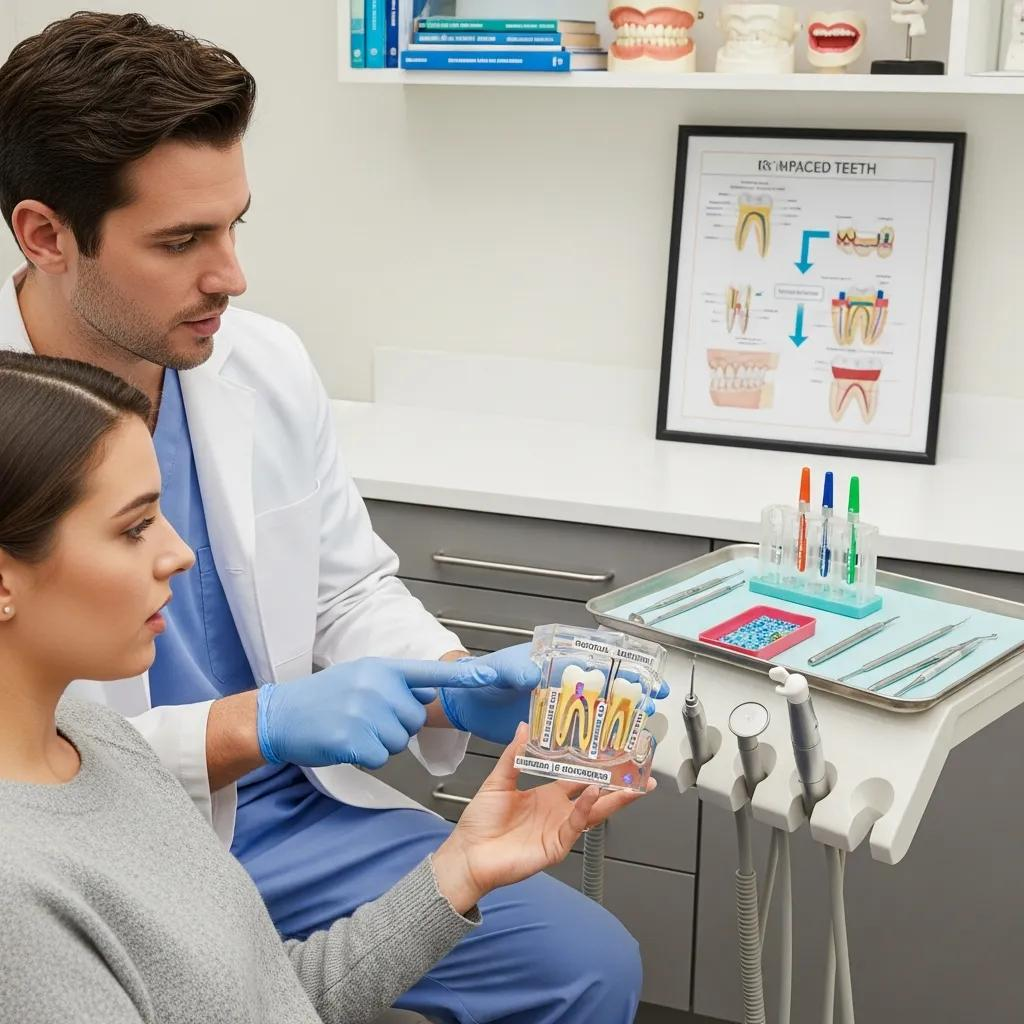
How Are Impacted Teeth Diagnosed at Bespoke Dental in Lutz, FL?
Diagnosis of impacted teeth begins with a focused dental history and clinical exam to identify symptoms, palpation findings, and visible soft-tissue changes that guide imaging choices and urgency. Imaging plays a central role: panoramic radiographs provide an overview of tooth position and relationships to neighboring teeth, while periapical films show finer bony detail; three-dimensional CBCT imaging is reserved for complex cases where the proximity of roots to nerves or unusual angulation affects surgical planning. A precise diagnosis allows clinicians to differentiate between simple eruption delay, pathological impaction, and infection-related issues and to recommend monitoring, orthodontic consultation, or extraction as appropriate. For patients in Lutz, FL seeking local evaluation, scheduling a clinical exam with appropriate imaging helps clarify whether intervention is needed and which modality—panoramic X-ray or CBCT—best informs safe treatment planning.
What Role Do X-rays and CBCT Scans Play in Diagnosis?
X-rays and CBCT scans provide complementary information: panoramic radiographs give a broad view of tooth orientation and jaw relationships, periapical films offer high-resolution views of roots and adjacent bone, and CBCT supplies three-dimensional detail when precise assessment of nerve proximity or impacted tooth angulation is required. CBCT imaging improves accuracy for surgical planning in complex impactions by mapping root morphology and bone contours relative to the inferior alveolar nerve or maxillary sinus, which reduces intraoperative surprises and informs conservative bone removal. However, CBCT is reserved for cases where 2-D imaging is insufficient because it involves higher radiation and cost; clinicians weigh benefit versus exposure when recommending CBCT. Understanding the strengths and limits of each modality helps patients appreciate why specific images are ordered.
Studies have further investigated the comparative accuracy of these imaging modalities, particularly concerning the critical proximity of impacted molars to vital nerves.
CBCT vs. Panoramic Radiography for Impacted Molar Diagnosis
Background:This study evaluated and determined the proximity of an impacted third mandibular molar (TMM) to the inferior alveolar canal (IAC) by using CBCT and digital panoramic radiography. The CBCT findings for the proximity of the TMM to inferior nerve canal used the outcomes of surgical findings as the standard of comparison. The result for CBCT radiographic diagnosis was 95%. Only 13.3% of the cases in which panoramic views showed the proximity of the TMM and the IAC were confirmed during surgery.
… of CBCT and Digital Panoramic Radiography with surgical findings to determine the proximity of an impacted third mandibular molar to the inferior alveolar nerve …, 2015
- Panoramic X-ray: Broad overview of dental arches and impaction position.
- Periapical X-ray: Detailed view of roots and localized bone changes.
- CBCT scan: 3D assessment for nerve proximity and complex surgical planning.
Choosing the right imaging modality depends on exam findings and surgical risk, with CBCT used selectively when it will change management.
How Does a Dental Exam Help Identify Impacted Teeth?
A dental exam identifies visual and tactile signs that suggest impaction, such as a partially erupted crown with an overlying gum flap, localized erythema, and tenderness on palpation, and these findings prioritize which radiographs to order. Exam maneuvers include inspection for drainage or pocketing, palpation for induration or fluctuance, assessment of occlusion and adjacent tooth health, and documentation of pain provocation by biting or percussion, which helps differentiate dental pain from other orofacial causes. The exam also evaluates mouth opening and muscular tenderness to detect functional impairment that may accompany infection or trismus. Together, these clinical observations guide imaging selection and triage urgency, leading into decisions about monitoring versus active intervention.
What Treatment Options Are Available for Impacted Teeth?
Treatment options for impacted teeth range from conservative monitoring to orthodontic exposure and surgical extraction, and selection depends on symptoms, pathology, patient age, and long-term dental plans. Monitoring is appropriate for asymptomatic impactions without pathology, with periodic clinical exams and radiographs to detect changes. Orthodontic exposure and bonding can reposition impacted canines and select teeth to preserve them for occlusion, while surgical extraction is indicated when pain, infection, damage to adjacent teeth, cyst formation, or orthodontic reasons outweigh the benefits of retention. Shared decision-making considers invasiveness, recovery time, and risk of complications so patients understand trade-offs before proceeding.
When Is Surgical Extraction Recommended?
Surgical extraction is recommended when an impacted tooth causes recurrent infection or pericoronitis, produces damage to adjacent teeth such as decay or resorption, forms or threatens cystic change, or interferes with orthodontic or prosthetic planning. The decision also considers symptom severity, patient medical status, and potential surgical risk; recurrent inflammation or radiographic signs of pathology typically push the balance toward removal. Clinicians discuss expected outcomes and possible complications with patients, explaining how extraction reduces infection risk and prevents progressive damage to neighboring structures. Understanding these criteria helps patients weigh the benefits of definitive surgical management against conservative approaches.
Are Orthodontic and Monitoring Approaches Effective Alternatives?
Orthodontic exposure and monitoring are effective alternatives when the impacted tooth has favorable position and the patient’s treatment goals include retention of the tooth for function or esthetics, as is often the case with impacted canines. Monitoring is suitable for asymptomatic, non-pathologic impactions where the risks of intervention exceed the current benefits; periodic imaging ensures timely detection of any developing pathology. Orthodontic exposure requires coordination between the surgical and orthodontic teams and can preserve natural dentition, but it may extend treatment duration and requires patient compliance. Choosing among these options depends on tooth type, angulation, adjacent structures, and the patient’s overall treatment plan.
Research further emphasizes the specific challenges and considerations involved in the diagnosis and treatment of impacted canines, highlighting their prevalence and the importance of tailored interventions.
Impacted Canine Diagnosis & Treatment Options
Impacted canines represent a significant dental anomaly with far-reaching implications for both orthodontic and surgical interventions [1]. These teeth, crucial for functional occlusion and aesthetic harmony, often fail to erupt into their correct positions, leading to a range of clinical challenges [4]. Maxillary canines, in particular, exhibit a higher propensity for impaction, with prevalence rates reported to be between 1–3% in various populations [9]. This prevalence underscores the necessity for dental professionals to possess a thorough understanding of the aetiology, diagnosis, and management of impacted canines to optimize patient outcomes [14].
… the success rates and effectiveness of surgical and orthodontic interventions for impacted canines: a systematic review of surgical and orthodontic interventions and a …, A Mancini, 2025
What Does the Impacted Tooth Extraction Procedure Involve?
An impacted tooth extraction involves preoperative assessment, anesthesia planning, surgical access through an incision, selective bone removal when necessary, possible tooth sectioning for safe removal, and closure with sutures, with attention to minimizing trauma and protecting adjacent structures. Pre-op planning includes reviewing imaging to map root form and nerve position, determining anesthesia or sedation needs, and discussing post-op expectations. During the procedure, the surgeon balances efficient removal with conservation of bone and soft tissue to promote uneventful healing. After surgery, clear home-care instructions and a follow-up visit support recovery and identify complications early.
How Is Wisdom Teeth Removal Performed at Bespoke Dental?
At Bespoke Dental in Lutz, FL, surgical planning begins with a clinical exam and appropriate imaging to determine the impaction type and nerve relationships, and IV sedation is offered as an option to improve patient comfort for those who prefer deeper relaxation during surgery. The clinical team employs standard surgical techniques—careful incisions, conservative bone removal, and tooth sectioning as required—aiming to remove the tooth while preserving surrounding tissues and minimizing postoperative discomfort. Intraoperative measures such as gentle handling of tissues and precise suturing reduce bleeding and enhance healing, and the care plan is individualized based on imaging findings and patient preferences. Scheduling a pre-op discussion helps patients understand sedation options and postoperative expectations before arriving for the procedure.
What Should Patients Expect During Recovery and Pain Management?
Recovery typically follows a predictable timeline: the first 24–72 hours often have the most pain and swelling as the inflammatory response peaks, while the first week sees steady improvement and sutures may be reviewed; by two weeks most soft-tissue healing is evident though deeper bone remodeling continues. Pain control strategies include prescribed or over-the-counter analgesics as recommended by the clinician, regular icing for the initial 24–48 hours, a soft diet to minimize chewing stress, and rest to allow healing. Patients are advised to avoid smoking, forceful rinsing, and strenuous activity in the immediate post-op period to reduce the risk of dry socket and delayed healing. If severe pain, increasing swelling, persistent bleeding, or fever occur beyond the expected timeline, patients should contact their dental provider for assessment.
Recovery timeline in brief:
- First 24–72 hours: peak swelling and discomfort; use ice and analgesics.
- First week: decreased pain and resumption of light activities; follow-up as scheduled.
- Two weeks and beyond: soft-tissue healing; complete bone remodeling over months.
What Are the Potential Complications and How Can They Be Managed?
Potential complications of impacted tooth extraction include localized infection, alveolar osteitis (dry socket), transient or, rarely, persistent nerve injury, and damage to adjacent teeth or sinus involvement in upper extractions; awareness and prevention strategies reduce their likelihood. Preventive steps include thorough preoperative imaging and planning, perioperative antiseptic measures, selective antibiotic use when indicated by systemic or local risk factors, and clear postoperative instructions to patients. Management pathways vary: infections typically respond to antibiotics and drainage if necessary; dry socket is treated with local medicated dressings and analgesics; nerve injuries are assessed and monitored with specialist referral as needed. Early recognition of complications and timely follow-up are critical to limit long-term sequelae and restore oral health.
Indeed, a comprehensive review of third molar surgery complications underscores the common risks and the various factors that can influence their incidence and management.
Impacted Third Molar Extraction Complications & Management
Third molar surgery is the most common procedure performed by oral and maxillofacial surgeons worldwide. This article addresses the incidence of specific complications and, where possible, offers a preventive or management strategy. Complications, such as pain, dry socket, swelling, paresthesia of the lingual or inferior alveolar nerve, bleeding, and infection are most common. Factors thought to influence the incidence of complications after third molar removal include age, gender, medical history, oral contraceptives, presence of pericoronitis, poor oral hygiene, smoking, type of impaction, relationship of third molar to the inferior alveolar nerve, surgical time, surgical technique, surgeon experience, use of perioperative antibiotics, use of topical antiseptics, use of intra-socket medications, and anaesthetic technique.
Complications after extraction of impacted third molars-literature review, EG Deliverska, 2016
ComplicationSigns to Watch ForLikelihoodManagementInfectionIncreasing swelling, fever, purulent drainageModerate if untreatedAntibiotics, drainage, follow-upDry Socket (Alveolar Osteitis)Severe radiating pain 3–5 days post-op, exposed boneModerateLocal dressing, analgesics, clinician careNerve InjuryNumbness or altered sensation in lip/tongueLow to rareMonitor, specialist referral if persistentAdjacent Tooth DamageNew sensitivity or looseningLowRestorative care or monitoring
This triage table helps patients know which signs require prompt dental attention and which warrant routine follow-up.
How Can Infection and Damage Be Prevented?
Preventing infection and tissue damage starts with careful pre-op assessment including targeted imaging, appropriate antiseptic technique during surgery, and educating patients on postoperative care like oral hygiene modifications and activity limitations. Selective antibiotic prophylaxis may be used for patients with significant infection or for specific surgical scenarios; clinicians decide based on current guidelines and individual risk factors. Surgical technique that minimizes bone removal and protects adjacent structures reduces iatrogenic damage, while clear written aftercare instructions and scheduled follow-up help clinicians detect early problems. Emphasizing prevention reduces the chance of complications and supports faster recovery.
When Should You Seek Immediate Care for Impacted Tooth Issues?
Immediate care is warranted for red-flag symptoms such as rapidly spreading facial swelling that threatens airway compromise, severe uncontrolled bleeding after an extraction, signs of systemic infection like high fever and rigors, or sudden onset of severe numbness that could indicate nerve compromise. If swelling increases rapidly or breathing/swallowing becomes difficult, emergency medical evaluation is required in addition to urgent dental intervention. For escalating pain unresponsive to recommended analgesics, persistent fever, or continuous bleeding beyond expected timelines, contacting your dental provider quickly enables assessment and expedient management. Prompt treatment of these serious signs prevents progression to life-threatening complications.
A short emergency-action checklist:
- Seek immediate care for airway compromise, rapidly spreading swelling, or severe systemic symptoms.
- Contact your dental provider for unmanageable pain, persistent bleeding, or high fever.
- Follow instructions for urgent evaluation and avoid delaying assessment.
Why Choose Bespoke Dental for Impacted Tooth Care in Lutz, FL?
Bespoke Dental offers patient-centered impacted tooth care that emphasizes personalized treatment planning, advanced imaging when indicated, and techniques to enhance comfort during surgical procedures. The practice highlights an expert clinical team including Dr. Tarik Elmohd and Dr. Michael Fabian, with services spanning general, cosmetic, and oral surgery focused on individualized outcomes. Patient comfort measures such as IV sedation are available for those who prefer deeper relaxation during procedures, and flexible financial options are offered to help patients access necessary care. For residents of Lutz, FL seeking diagnosis and treatment of impacted wisdom teeth, arranging an exam with imaging when appropriate provides a clear plan tailored to clinical findings and patient preferences.
What Expertise Does Dr. Tarik Elmohd Bring to Oral Surgery?
Dr. Tarik Elmohd leads the Bespoke Dental team with a clinical role in oral surgery, contributing specific expertise in managing impacted teeth and related surgical procedures as described in local practice information. His involvement supports advanced surgical capability for complex impactions, enabling careful planning and execution to protect adjacent structures and reduce complication risk. Patients benefit from having surgical expertise integrated within a broader dental practice, which streamlines diagnosis, imaging review, and coordinated postoperative care. This clinical focus assists in delivering predictable outcomes for impacted tooth removal.
How Do Personalized Care and Advanced Technology Improve Outcomes?
Personalized care at Bespoke Dental pairs detailed clinical exams with appropriate imaging—using panoramic X-rays and selective CBCT scans—to create individualized surgical plans that minimize unnecessary invasiveness and better protect nerves and adjacent teeth. Advanced technology aids accurate diagnosis and surgical mapping, while patient-centered measures such as IV sedation enhance perioperative comfort and reduce anxiety, contributing to smoother procedures and recovery. Flexible financial options help align treatment choices with patient circumstances, and scheduled follow-up visits ensure complications are identified and managed quickly. Together, personalized planning and modern imaging support safer extractions and improved recovery experiences for patients in the Lutz area.