
Signs of an Impacted Tooth You Shouldn't Ignore
An impacted tooth is one that fails to fully emerge through the gum line because it is blocked by adjacent teeth, bone, or an abnormal eruption path, and recognizing early signs of impaction reduces the risk of infection, damage to nearby teeth, and more invasive treatment.
This article explains what causes impacted teeth, how to spot the most common signs—especially for impacted wisdom teeth—and when prompt evaluation is necessary to prevent complications. You will learn the typical symptom pattern for impacted tooth pain, how swelling or bad breath can signal pericoronal infection, what imaging and exams reveal, and the standard treatment pathways including extraction, sedation choices, and implant planning. Practical sections cover immediate at-home relief, red flags that require urgent care, and a clear aftercare checklist to support healing following extraction. Throughout, the content integrates current terms like pericoronitis, trismus, and types of impaction while offering actionable steps to help you know when to see a dental professional.
What Is an Impacted Tooth and How Does It Occur?
An impacted tooth is an unerupted tooth that remains partially or completely below the gum line because its eruption path is blocked or misaligned, often producing local inflammation or functional problems. Mechanical factors such as lack of space in the dental arch, abnormal angulation of the developing tooth, or obstruction by neighboring teeth or dense alveolar bone explain why eruption fails; developmental contributors include jaw size, tooth size, and genetic variation in eruption timing. Understanding these causes clarifies why impacted teeth are common in late adolescence and early adulthood, particularly for third molars and certain canines, and why orientation of the tooth (its angulation relative to neighboring structures) affects both symptoms and extraction complexity. The table below summarizes common impaction orientations, their typical frequency, and principal causes to help readers recognize patterns that can inform diagnosis.
What Are the Different Types of Tooth Impaction?
Vertical, mesioangular, horizontal, and distoangular orientations each describe how an impacted tooth sits relative to the dental arch and adjacent teeth, and these positions influence symptom patterns and extraction complexity. Vertical impactions may remain asymptomatic or cause mild pericoronal irritation, while mesioangular and horizontal impactions frequently press on neighboring tooth roots, producing referred pain and increased risk of decay in adjacent enamel. Horizontal impactions are often the most technically challenging to remove because the crown lies against the side of a neighboring tooth and may require sectioning of the tooth during extraction; distoangular impactions, while less common, can complicate access and increase the likelihood of extended healing. Recognizing the impaction type on a radiograph guides whether monitoring, surgical extraction, or referral to an oral surgeon is the most appropriate next step.
How Does Dental Crowding and Genetics Cause Tooth Impaction?
Dental crowding limits eruption space when tooth size exceeds available arch length, forcing developing teeth to take an aberrant path or remain unerupted, and heredity commonly determines jaw size and tooth dimensions that predispose families to impaction. Genetic patterns influence timing of growth and eruption, so siblings or parents with a history of impacted third molars or canine impaction often signal a higher individual risk; epidemiologic data show higher rates of third molar impaction in populations with smaller mandibular arches. Growth patterns during adolescence—when wisdom teeth commonly attempt to erupt—interact with late-stage orthodontic changes and further influence whether teeth can erupt normally. Understanding the interplay of crowding and genetic predisposition helps patients anticipate the need for monitoring or early intervention during late teen and young adult years.
What Are the Common Symptoms of an Impacted Wisdom Tooth?
An impacted wisdom tooth commonly manifests through a consistent cluster of symptoms that reflect local tissue inflammation, mechanical pressure, and bacterial overgrowth around a partially erupted crown. Typical signs include localized pain that may radiate to the ear or jaw, swollen or tender gum tissue around the back molar, persistent bad breath due to trapped debris, and difficulty fully opening the mouth (trismus) when inflammation involves the muscles of mastication. These symptoms can be intermittent early on and become recurrent or progressively worse if an infection like pericoronitis develops; recognizing the pattern early supports timely diagnostic imaging and treatment.
Common signs that suggest an impacted wisdom tooth:
- Localized pain or aching: Dull, persistent pain near the back molar that may radiate to the jaw, ear, or temple.
- Gum swelling or tenderness: Inflamed tissue near a partially erupted tooth, often worse when chewing.
- Bad breath or unpleasant taste: Halitosis caused by trapped food, plaque, and bacterial colonization under a pericoronal flap.
- Limited mouth opening (trismus): Stiffness of jaw muscles from local inflammation or infection.
These symptomatic clues point toward probable impaction and should prompt diagnostic imaging or clinical evaluation to confirm the diagnosis and plan appropriate care.
How Does Impacted Tooth Pain Present and How Can It Be Relieved?
Pain from an impacted tooth is often localized and persistent, described as a deep aching that can refer to adjacent structures such as the jaw, ear, or temporomandibular region, and it typically intensifies with biting or pressure. Short-term relief measures include over-the-counter analgesics taken per label directions, cold compresses applied externally to reduce swelling, and gentle rinsing with warm salt water to flush debris when infection is not severe; these measures offer symptom control but do not resolve the underlying impaction. Patients should avoid placing aspirin directly on exposed gums and should not attempt to forcefully manipulate impacted tissue, since that can worsen inflammation or spread infection. Persistent, worsening, or systemic symptoms such as fever or difficulty breathing call for urgent professional assessment rather than continued home treatment.
What Signs Indicate Gum Swelling, Bad Breath, or Jaw Stiffness from Impaction?
Gum swelling and a persistent bad taste often reflect pericoronal inflammation or localized infection when a partly erupted tooth traps food and bacteria beneath the gum flap, while progressive jaw stiffness (trismus) suggests extension of inflammation into the masticatory muscles. Mild irritation presents as tender, slightly swollen gum tissue around the crown and can often be managed conservatively with improved hygiene and warm rinses, but the same signs accompanied by fever, spreading redness, or increasing pain likely indicate bacterial infection requiring professional treatment. Bad breath in this context stems from anaerobic bacterial activity and debris accumulation in the pericoronal pocket, and it tends to persist until the source is cleaned or removed. Distinguishing between minor irritation and signs of infection helps determine whether home care or prompt dental intervention is the correct next step.
What Complications Can Arise from Ignoring an Impacted Tooth?
Untreated impacted teeth can lead to several progressive complications that affect both local oral structures and, in some cases, overall health; early identification reduces the likelihood of these outcomes. Left unmanaged, impactions can develop recurrent pericoronitis (localized gum infection), create dental cysts that resorb adjacent bone, accelerate decay in neighboring teeth due to trapped plaque, and cause periodontal breakdown around adjacent molars. In rare cases infection can spread to deeper facial spaces or contribute to systemic inflammatory burden if not controlled, emphasizing why monitoring and timely treatment matter.
Key risks of untreated impacted teeth include:
- Recurrent infection (pericoronitis): Repeated inflammation around the tooth leading to pain and swelling.
- Cyst or tumor formation: Fluid-filled cysts may develop in association with an unerupted tooth, causing bone loss.
- Damage to adjacent teeth: Pressure and bacterial spread can produce caries or root resorption on neighboring teeth.
How Can Untreated Impacted Teeth Lead to Infections, Cysts, or Damage to Adjacent Teeth?
Bacterial colonization under the soft-tissue flap of a partially erupted tooth creates a persistent infectious nidus that can cause pericoronitis; the same bacterial and inflammatory processes promote cystic degeneration of follicular tissue and can erode adjacent alveolar bone. Accumulated plaque and food debris predispose the adjacent tooth to caries and periodontal inflammation, and ongoing pressure from an impacted crown may induce root resorption or compromise the periodontal attachment of neighboring teeth. Imaging studies routinely reveal the relationship of an impacted tooth to surrounding structures, and early removal or careful monitoring prevents progression from localized soft-tissue infection to more extensive bony involvement. Recognizing these pathological mechanisms helps patients understand why conservative home care is only a temporary measure in many cases.
What Is Pericoronitis and Why Is Early Treatment Important?
Pericoronitis is the inflammation and infection of the soft tissue (pericoronal flap) surrounding a partially erupted tooth, commonly seen with lower third molars, and it presents with pain, swelling, and sometimes systemic symptoms like fever when severe. Early treatment focuses on reducing inflammation and bacterial load with antiseptic rinses, debridement or irrigation, and antibiotics when indicated; persistent or recurrent pericoronitis usually requires definitive management such as surgical removal of the offending tooth or operculectomy in select cases. Prompt attention prevents spread of infection into deeper facial spaces, reduces the chance of chronic follicular pathology, and limits damage to adjacent dentition. Timely intervention therefore lowers the risk of complications and simplifies definitive treatment planning.
When Should You See a Dentist for an Impacted Tooth?
You should seek dental evaluation when symptoms are persistent, progressively worse, or if any red-flag signs appear; urgent assessment is warranted for severe pain, swelling that affects breathing or swallowing, fever, or signs of spreading infection. For non-urgent but concerning symptoms—recurrent pain, recurrent swelling, bad breath that does not respond to hygiene, or difficulty chewing—schedule a clinical examination and imaging to determine impaction status and treatment options. Diagnostic assessment typically includes a focused oral exam and radiographic imaging such as periapical or panoramic X-rays, with cone-beam CT reserved for complex cases where nerve location or root anatomy affects surgical planning. If you are local to Lutz, FL, Bespoke Dental provides clinical exams, radiographic diagnostics, and oral surgery consultation options to evaluate impacted teeth and plan care with an oral surgery specialist when indicated.
When to see a dentist — a short checklist:
- Severe or escalating pain: Unresponsive to OTC analgesics or interfering with sleep.
- Spreading swelling or fever: Signs of systemic infection requiring urgent care.
- Recurrent localized swelling or bad breath: Persistent pericoronal symptoms that recur frequently.
This checklist clarifies when immediate attention is needed and leads into a description of the imaging tools used to confirm impaction and guide treatment.
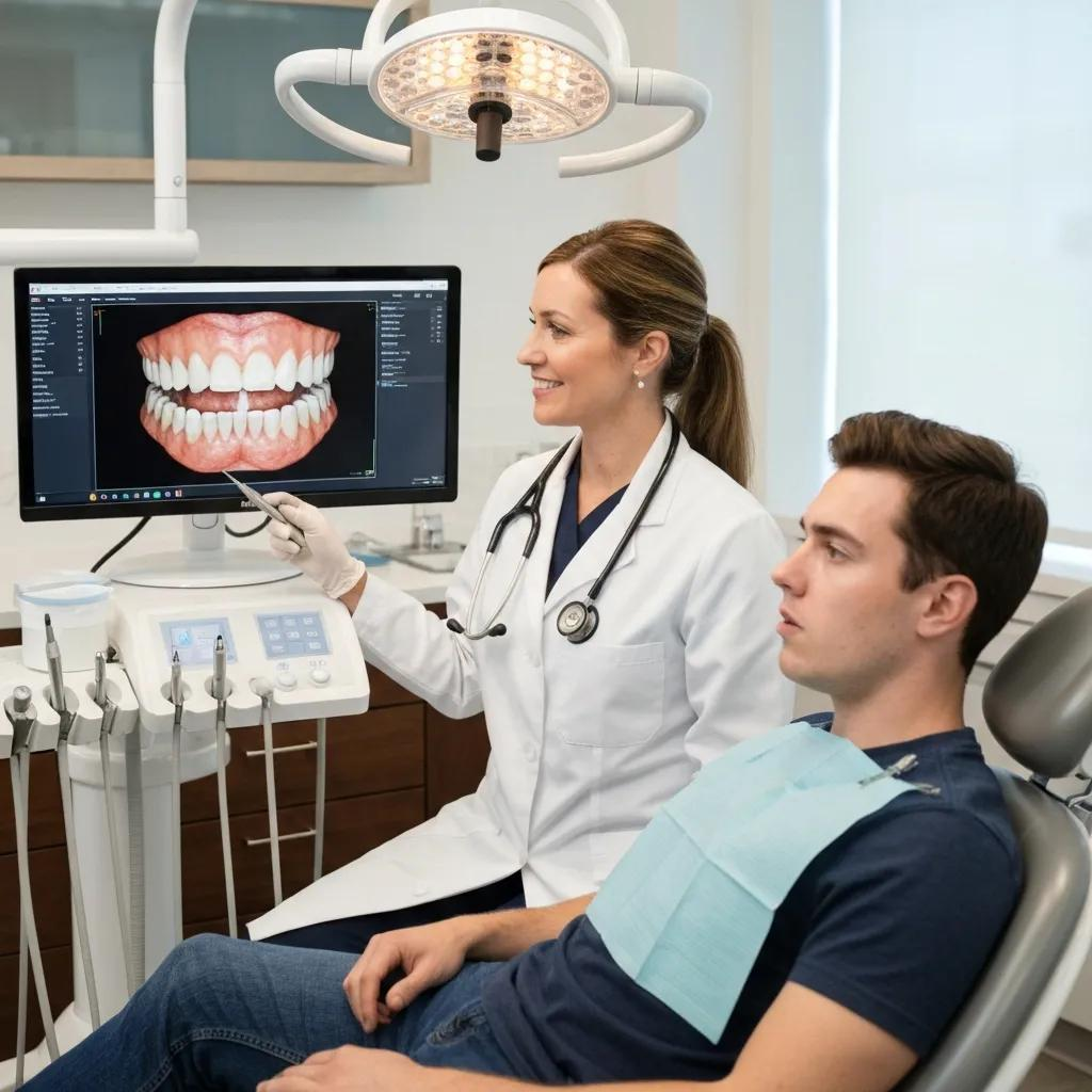
What Diagnostic Tools Are Used to Identify Impacted Teeth?
Clinical exam combined with radiographic imaging forms the backbone of impaction diagnosis; panoramic radiographs show overall tooth position and relationships within the jaw, while periapical films provide detail on adjacent roots and local pathology. Cone-beam computed tomography (CBCT) gives three-dimensional detail for complex impactions, clarifying proximity to the inferior alveolar nerve, sinus floor, or adjacent roots to optimize surgical planning and reduce procedural risk. These imaging modalities inform whether monitoring, simple extraction, or referral to an oral surgeon is the safest course and also help in planning implant placement if extraction is followed by tooth replacement. Understanding what each tool reveals helps patients appreciate why certain images are recommended before proceeding with definitive treatment.
Further research emphasizes the critical role of advanced imaging like CBCT in precisely mapping anatomical structures for safer surgical interventions.
CBCT Imaging for Impacted Tooth Assessment
The objective of this study was to quantify the ability of cone beam computed tomography (CBCT) to measure the length of the mental nerve loop, the length and diameter of the incisive nerve canals, and the incisive canal path.
Retrospective study of the anterior loop of the inferior alveolar nerve and the incisive canal using cone beam computed tomography, BS Sotto-Maior, 2013
How Does Dr. Tarik Elmohd’s Oral Surgery Expertise Support Impacted Tooth Treatment?
Board-certified oral surgeons play a key role in evaluating and managing complex impactions, and Dr. Tarik Elmohd (board-certified oral surgeon) provides specialist assessment and surgical care for cases that require advanced techniques, sedation, or careful nerve-sparing planning. Specialist involvement matters for horizontal impactions, proximity to nerve structures, or patients with medical complexities where IV sedation or surgical access techniques improve outcomes and comfort. Bespoke Dental collaborates across general dentistry and oral surgery to ensure that diagnostic findings translate into safe, individualized treatment plans, and specialist consultation reduces the risk of unexpected intraoperative challenges. This interdisciplinary approach helps patients receive both accurate diagnosis and an appropriate procedural plan tailored to their anatomy and comfort needs.
What Are the Treatment Options for Impacted Teeth at Bespoke Dental?
Treatment options range from conservative monitoring to surgical extraction, with choices guided by symptoms, impaction type, and patient-specific factors like age and systemic health; at Bespoke Dental these pathways include oral surgery for wisdom tooth extraction, IV sedation when appropriate, and implant planning for replacement when extraction is definitive. Indications for extraction include recurrent infection, damage to adjacent teeth, cyst formation, or significant pain; monitoring with periodic imaging may be reasonable for asymptomatic, deeply impacted teeth without pathology. Sedation options available include local anesthesia with adjunctive nitrous oxide for mild anxiety, and IV sedation for patients requiring deeper anxiolysis or when complex surgical access is anticipated. For patients who elect extraction and desire tooth replacement, dental implant planning is integrated into the post-extraction workflow to restore function and aesthetics as indicated.
How Is Wisdom Tooth Extraction Performed and What Sedation Options Are Available?
Wisdom tooth extraction typically begins with a thorough exam and imaging to map tooth position; the procedure involves local anesthesia to numb the area, careful elevation and removal of bone overlying the tooth if necessary, and sectioning of the tooth when orientation requires it, followed by suturing of the surgical site. Nitrous oxide or oral sedatives may be offered for patients with moderate anxiety, while IV sedation is available for deeper sedation needs and is particularly useful when multiple extractions or complex impactions are planned to improve patient comfort and procedural efficiency. Postoperative instructions include pain management and hygiene measures to protect the clot and promote healing; selecting the sedation option depends on medical history, anxiety level, and the anticipated surgical complexity. Discussing these choices with the provider ensures individualized care and clarity on what to expect during and after the procedure.
When Are Dental Implants Recommended After Tooth Extraction?
Dental implants are recommended when replacing a missing tooth will restore chewing function and aesthetics and when the patient has adequate bone volume or can undergo grafting to support osseointegration; timing may be immediate, early, or delayed depending on infection status and bone quality. Immediate implant placement may be possible in sites free of active infection and with sufficient native bone to stabilize the implant, whereas chronic infection, significant bone loss, or the need for alveolar ridge preservation often warrant delayed placement following healing and possible grafting. Implant candidacy also considers systemic health, smoking status, and patient goals; when implants are planned, the oral surgery and restorative teams collaborate on prosthetic design and timing. Clear communication between the surgical and restorative clinicians optimizes functional and aesthetic outcomes.
How Should You Care for Your Mouth After Impacted Tooth Extraction?
After extraction, initial actions support clot formation, reduce bleeding, and lower infection risk; immediate care involves gentle gauze pressure, avoiding rinsing or spitting for the first 24 hours, and using cold packs externally to limit swelling. Pain management typically combines scheduled analgesics per provider guidance and adjunctive measures like ice for the first 48 hours, while oral hygiene should continue with careful brushing away from the surgical site and gentle saline rinses after 24 hours to keep the area clean without disturbing the clot. Activity adjustments include rest and avoiding heavy lifting or vigorous exercise for several days to minimize rebound bleeding and reduce the risk of dry socket; dietary progression begins with soft, cool foods before slowly returning to a normal diet as tolerated. The table below summarizes practical aftercare actions by category to serve as a quick-reference guide for patients.
Care CategoryIssue AddressedRecommended ActionBleeding controlHemostasisApply firm gauze for 30–60 minutes; replace as neededPain managementDiscomfort controlUse prescribed or OTC analgesics on schedule; ice intermittentlyOral hygieneInfection preventionGentle brushing; saline rinses starting 24 hours post-opDiet & activityProtect clotEat soft foods; avoid straws, smoking, heavy exertion
This table consolidates essential aftercare steps and leads into specific recommendations for medication use and diet progression to support uncomplicated healing.What Are the Best Practices for Pain Management and Oral Hygiene Post-Extraction?
Effective pain control relies on a combination of appropriate analgesics taken at recommended intervals, local measures such as cold packs during the first 48 hours to reduce swelling, and avoiding actions that disturb the clot like forceful rinsing or sucking on straws. When prescription analgesics are provided, follow dosing instructions closely and discuss any contraindications with your provider; for many patients, scheduled nonsteroidal anti-inflammatory drugs plus acetaminophen provides reliable control. Oral hygiene should focus on keeping the rest of the mouth clean while protecting the surgical site: brush gently away from the socket, start warm saline rinses after the first day, and avoid alcohol-based mouthwashes until healing is established. Contact the clinic if pain escalates after an initial improvement, if there is a foul odor with increasing pain (possible dry socket), or if you experience fever, since these signs require prompt assessment.
What Diet and Activity Adjustments Support Healing After Surgery?
Diet in the first 24–48 hours should emphasize cool, soft foods such as applesauce, yogurt, smoothies (consumed without a straw), and pureed soups, progressing to warm soft foods as comfort allows over the next week; crunchy, spicy, or chewy items should be avoided until the surgical site demonstrates healing. Activity recommendations prioritize rest for the first 48 hours and avoidance of heavy lifting, bending, or intense aerobic exercise that might increase blood pressure and provoke bleeding or clot disruption. Smoking, use of straws, or vigorous rinsing are specific behaviors to avoid because they risk creating negative pressure and dislodging the clot, leading to dry socket and delayed healing. Returning to normal diet and activity depends on individual healing and comfort; routine follow-up with your provider ensures the site is healing and clarifies when progressive activity is safe.
For patients in the Lutz, FL area seeking evaluation or treatment for an impacted tooth, Bespoke Dental offers diagnostics, oral surgery services (including wisdom teeth and extractions), IV sedation for patient comfort, and restorative planning such as dental implant coordination with the surgical team. To schedule an evaluation or oral surgery consultation, contact Bespoke Dental at (813) 807-4902; the practice’s team includes Dr. Tarik Elmohd (board-certified oral surgeon) and Dr. Michael Fabian (licensed dentist), who work together to provide advanced technology and personalized patient care when managing impacted teeth.